 ALERT
ALERT * * * Important Update for iLab Users * * *
New Support System Now Live: We are pleased to share that the new iLab Help Desk platform is now live as of January 14th, 2026.
The ‘Help’ link in iLab now opens an email through the new ticketing system. For details, please visit our Help Site article: Submit and Track Support Requests in the Portal.
Upcoming Holiday Closure: In observance of a holiday, Agilent CrossLab/iLab Operations Software Support Help Desk will be closed during U.S. hours on Monday, January 19th, 2026. During this time, support in the U.S. and Canada will be unavailable. APAC and EU Support will remain open as normal. For urgent matters, please add "Urgent" to your ticket/email subject or press "1" when prompted to escalate a call on the iLab Support phone, and we will prioritize those requests first.

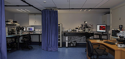
The Microscopy and Imaging Facility at TBSI is integrating advances in Microscopy with Computer Imaging. The services of this facility are available to all researchers, both internal and external to the University. The instruments are designed for analysis of multiple dyes on fixed or live samples (e.g. multicolor immunofluorescence, photoactivation, colocalisation studies FRET & FRAP). It has been developed to facilitate research in the biological sciences through the use of state of the art imaging systems.
We have a Leica SP8 gated STED with 5 detectors and a white laser. It also has a tandem scanner that allows you to take 24 frames a second. We also have a standard Leica SP8 confocal with 3 detectors.
We have two systems for epifluorescence imaging, an Olympus BX51 upright microscope with four colour LED and an IX81 inverted microscope for long working distance.
Along with the equipment we provide application support, including experimental setup, sample preparation advice and troubleshooting along with software analysis of the results using Imaris software from Bitplane.
Dr Barry Moran
Director of Research Technology
01-8962761
https://www.tcd.ie/Biochemistry/research/facilities/microscopy-imaging
Facility is located in B2.46 TBSI
| Name | Role | Phone | Location | |
|---|---|---|---|---|
| Dr Barry Moran |
Interim Lead
|
01-8962761
|
barry.moran@tcd.ie
|
TBSI 3.09
|